What is craniosynostosis?

Craniosynostosis is the premature and abnormal fusion or closure of one or more of the cranial suture lines that form the skull. Premature suture closure results in an abnormal head shape and abnormal bone growth pattern. If uncorrected, it can lead to continued abnormal growth of the skull and abnormalities in the shape and symmetry of the face. On occasion it can lead to increased intracranial pressure.

The normal newborn skull is composed of the frontal, parietal, temporal, sphenoid and occipital bones. Having these gaps (described as open sutures) allows for continuous separation of the skull bones during fetal brain growth and allows for molding of the head to facilitate passage through the birth canal. After birth, these open sutures also allow for continued growth and expansion of the head during the first few years of life. Head growth is driven by growth of the brain. If these sutures close prematurely growth of the skull is restricted in one or more directions. The skull attempts to compensate for this and has an altered growth pattern which leads to a significant deformity of the skull.

CONTACT US

For questions or to make an appointment, please call the cleft and craniofacial clinic at 612-813-6888.

An image of a skull displaying different Craniosynostosis Sutures

What are the different types of craniosynostosis?

Scaphocephaly or boat shaped skull occurs following fusion of the sagittal suture, which runs from the front of the head to the back of the head, right down the middle. Often, a ridge is seen or felt along this suture line. Sometimes, the fontanel, or soft spot, is absent or closed. The skull is significantly expanded in the anterior–‐‑posterior direction with growth restriction in the transverse direction. This gives the appearance of a long and narrow skull. The incidence of scaphocephaly is one in 2,000 births. It is the most common form of craniosynostosis.

An image of a skull demonstrating Craniosynostosis Sagittal

Brachycephaly or short skull occurs secondary to premature fusion of both coronal sutures. The skull is short in anterior–‐‑posterior growth. The skull grows vertically and laterally leading to a high and wide skull shape.

An image of a skull demonstrating Craniosynostosis Metopic

Unilateral coronal synostosis results in this short high skull on one side. The affected side of the skull tends to be wide in this condition with a ridge felt running over the top of the front side of the head. The eyebrow on the affected side is pulled upward, causing the eye opening to appear wider than the normal side. When viewed from above, the forehead on the affected side is recessed, or further back than the other side. The root, or top of the nose, is deviated toward the side of the fused suture causing the tip of the nose to look deviated towards the opposite side. The nose looks tilted. The incidence of unilateral coronal synostosis is estimated to be about one in 3500 births.

An image of craniosynostosis coronal on the left side of the skull

Fusion of the metopic suture results in trigonocephaly or triangular skull. The metopic suture runs from the top of the head, from the soft spot, down the center of the forehead to the nose. It normally begins closing in infancy. In cases of trigonocephaly, the forehead is narrowed and pointed and there is a ridge running down the middle of the forehead to the top of the nose. The eyes are usually spaced too close together. When viewed from above the forehead has a triangular shape, like the bow of a boat. The incidence of trigonocephaly is estimated at 1 in 2,500 to 1 in 3,500 births. It is sometimes difficult to determine if a child has trigonocephaly or just a small ridge running down the forehead. Many children may just have a ridge running down the center of their forehead suggesting that the metopic suture has closed a little bit early, without causing true trigonocephaly. Only those children who have a real triangular shape to the forehead and closely spaced eyes need treatment. Children with an isolated ridge running down their foreheads do not require surgery.

An image of a skull displaying Craniosynostosis Bicoronal

Lambdoid synostosis is a rare form of craniosynostosis that leads to flattening of the back part of the skull on one side. The ear is pulled backward, toward the fused suture. Another rare type of craniosynostosis is oxycephaly, also known as turricephaly or towering skull. It is a combination of multiple sutures fusing early. In Kleeblattschadel or cloverleaf skull, all cranial sutures except the metopic and squamosal sutures fuse prematurely giving the skull the appearance of a cloverleaf.

What causes craniosynostosis?

Craniosynostosis is a rare birth anomaly and is present in about three to five cases per 10,000 live births. The most common type is sagittal synostosis, followed by metopic synostosis, followed by unilateral coronal craniosynostosis and lambdoid synostosis is the rarest. Associated syndromic conditions such as Apert syndrome or Crouzon syndrome are quite rare. Doctors and scientists are not sure of the exact cause of craniosynostosis. Some think it is a genetic condition, while others think the premature closure is caused by the abnormal suture itself and still others believe that it is an abnormality of the dura which is the covering overlying the brain which causes premature fusion of the suture.

When and why is treatment required?

The indications for surgical intervention include the following:

  • Prevention of elevated intracranial pressure that can occur in some patients with uncorrected craniosynostosis.
  • Prevention of progression of the skull deformity.
  • Prevention of progression of the facial deformity.
  • Optimization of growth potential of the brain.

The general consensus currently holds that the best time for surgical repair is between three and six months of age. A delay in surgery typically leads to continuation of the deformity of the craniofacial skeleton. In addition, if increased intracranial pressure is present, this will adversely affect the development of the child.

How is craniosynostosis treated?

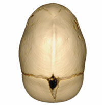
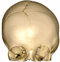
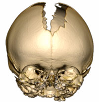
A three‑dimensional CT scan is obtained in suspected cases of craniosynostosis to confirm the diagnosis and to aid in surgical planning.

Craniosynostosis is treated with surgical correction. A team of surgeons consisting of a neurosurgeon and a craniofacial surgeon performs the surgery. The surgery can be done with an open technique or with a minimally-invasive endoscopic approach, depending on the condition and timing of repair. At Children’s Minnesota, the Neurosurgeon and Craniofacial surgeon collaborate before the procedure and then work together for the entire duration of the surgery.

The child is cared for in the pediatric intensive care unit for a few days after surgery. Most children resume a regular diet soon after the procedure and recovery quickly. It is rare but on occasion a child benefits from a secondary corrective surgical procedure at a later date. Occasionally a molding helmet is also helpful after surgical correction, but this is rarely needed. The child is then followed in the multidisciplinary Craniosynostosis Clinic to monitor growth and development.